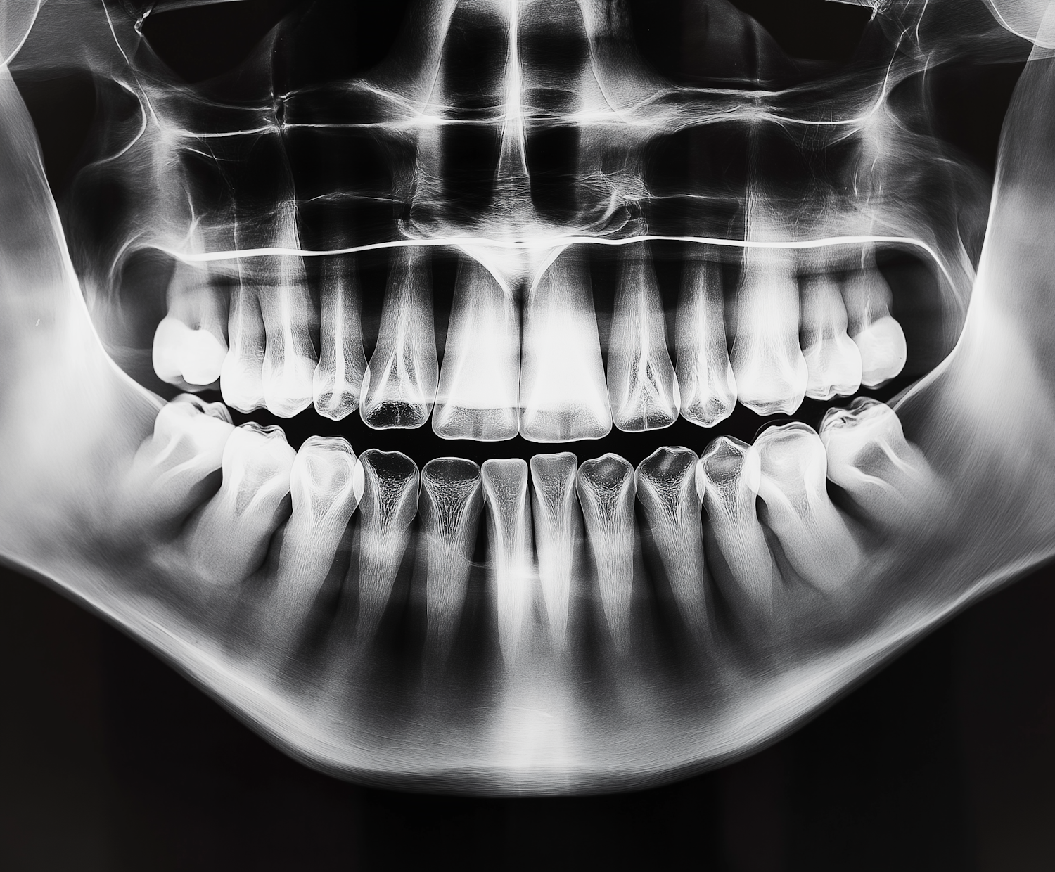
Human Jaw X-Ray
The image is a detailed X-ray of a human jaw showing both upper and lower sets of teeth. The X-ray is monochrome, with stark contrasts between the bright whites of the teeth and the darker shades outlining the jawbones and surrounding structures. There is a distinctive clarity in the depiction of the individual teeth, each displaying unique shapes and root structures; some molars also show fillings. The X-ray imaging technique allows for a detailed examination of the dental and skeletal structure of the mouth, often used in medical diagnostics to assess dental health and alignment, as well as to identify any dental abnormalities such as cavities or impacted teeth. This type of image is utilized in dental and medical fields, reflecting its clinical and diagnostic relevance.
Colors
Categories
Views
Downloads
Collected